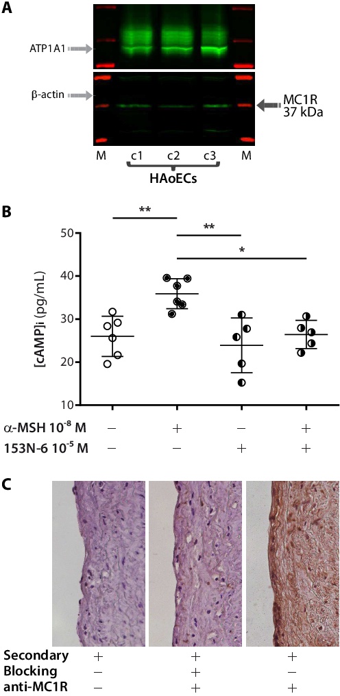
Fig. 1. HAoECs express a functional MC1R. (A) Immunoblot analysis of membrane extracts showed that all three studied primary cells express MC1R on the plasma membrane. An anti-ATPase Na+/K+ transporting subunit alpha 1 (ATP1A1) antibody was used as membrane-specific loading control (upper light grey arrow). Absence of a β-actin immunoreactive band (lower light grey arrow) excluded contamination with cytoplasmic proteins in this preparation. The 37 kDa MC1R-specific immunoreactive band is indicated by a grey arrow. Lanes are: M, molecular weight marker; C+, HEK293 cells transiently transfected with the MC1R full-length cDNA (positive control); c1, c2, c3, primary HAoECs from ECACC, Lonza, and Promocell, respectively. (B) Intracellular cAMP concentrations were measured in confluent HAoECs after treatment with α-MSH for 5 min, with or without the MC1R-selective α-MSH antagonist 153N-6. Results are shown as scatter dot plots with mean ± SD (n = 5-6 per treatment group). Statistical significance of differences was assessed by one-way ANOVA [F(3,18) = 7.900, p=0.0014] followed by Tukey's post-hoc test (*p<0.05, **p<0.01). (C) Immunohistochemical detection of MC1R in a normal human aorta specimen (10×) confirmed that HAoECs express the receptor in vivo. To control for staining specificity, we used secondary antibody alone (left), anti-MC1R antibody pre-adsorbed with the specific blocking peptide and secondary antibody (centre), and anti-MC1R antibody with secondary antibody (right): only the latter showed an intense staining.